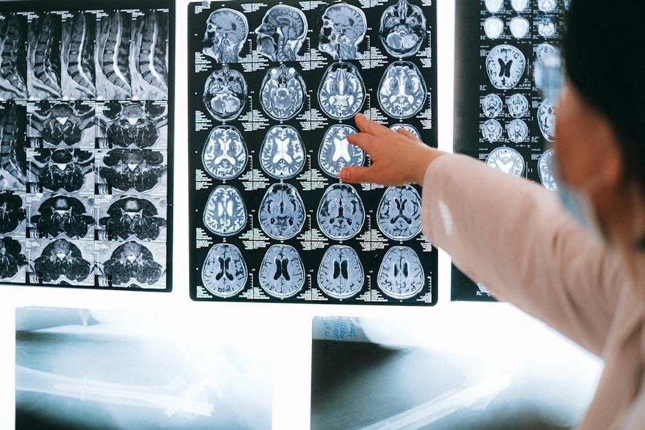
The humerus‚ the longest bone in the upper limb‚ connects the shoulder and elbow‚ enabling diverse movements. Its anatomical structure includes a shaft‚ epicondyles‚ and vital articulation points.
Overview of the Humerus Bone
The humerus is the longest and largest bone in the upper limb‚ extending from the shoulder to the elbow. It serves as the primary structural and functional component of the arm‚ enabling a wide range of movements. The bone is divided into three main sections: the proximal end‚ shaft‚ and distal end. The proximal end features the head and greater tubercle‚ which facilitate shoulder articulation. The shaft provides attachment points for muscles‚ while the distal end includes condyles and epicondyles‚ essential for elbow function. Its anatomical features‚ such as the deltoid tuberosity and radial groove‚ highlight its role in both movement and stability‚ making it vital for upper limb mobility and prone to fractures in certain cases.
Importance of Humerus in Upper Limb Anatomy
The humerus is a cornerstone of upper limb anatomy‚ providing structural support and enabling complex movements. It articulates with the scapula at the shoulder and the radius and ulna at the elbow‚ facilitating a wide range of motions. As the longest bone in the upper limb‚ it serves as a vital attachment point for numerous muscles‚ tendons‚ and ligaments‚ essential for arm mobility and stability. Its unique anatomy‚ including the deltoid tuberosity and epicondyles‚ allows for the distribution of forces during activities‚ making it indispensable for both functional and mechanical roles in the upper extremity.
Structure of the Humerus
The humerus consists of a proximal end‚ shaft‚ and distal end‚ each with distinct anatomical features that facilitate movement and support the upper limb’s functional requirements.
Proximal End of the Humerus
The proximal end of the humerus includes the head‚ anatomical neck‚ and greater tubercle. The head articulates with the glenoid cavity of the scapula‚ forming the shoulder joint. The anatomical neck‚ located just below the head‚ is a groove that houses the shoulder capsule and ligaments. The greater tubercle‚ a bony prominence‚ serves as an attachment site for muscles like the supraspinatus‚ infraspinatus‚ and teres minor. These muscles contribute to shoulder stabilization and movement. This region is crucial for upper limb mobility and is often susceptible to fractures‚ particularly in older adults. Its intricate structure supports both stability and a wide range of motion in the shoulder joint.
Shaft of the Humerus
The shaft of the humerus is the long‚ cylindrical portion between the proximal and distal ends. It provides attachment points for muscles and ligaments‚ facilitating movement and stability. The deltoid tuberosity‚ a roughened area on the lateral side‚ serves as the insertion point for the deltoid muscle. Additionally‚ the radial groove‚ located posteriorly‚ houses the radial nerve and profunda brachii artery. The shaft’s structure allows for efficient transmission of forces from the shoulder to the elbow‚ enabling a wide range of upper limb movements. Its robust design supports both weight-bearing and dynamic activities‚ making it a critical component of the upper limb’s functional anatomy.
Distal End of the Humerus
The distal end of the humerus forms the elbow joint‚ articulating with the radius and ulna bones of the forearm. It features two condyles—trochlea and capitulum—that facilitate flexion and extension of the elbow. The medial and lateral epicondyles project from the condyles‚ serving as attachment points for muscles controlling forearm and wrist movements. The supracondylar ridge‚ a bony prominence above the condyles‚ adds strength to the distal humerus. This region is crucial for both mobility and stability‚ enabling precise movements of the elbow and forearm. Its intricate design supports daily activities‚ making it a vital component of upper limb anatomy and function.
Proximal Humerus Anatomy
The proximal humerus includes the head‚ anatomical neck‚ and greater tubercle‚ forming the shoulder joint. It enables articulation with the scapula and muscle attachments for upper limb movement.
Head of the Humerus
The head of the humerus is a rounded‚ smooth structure located at the proximal end of the bone. It articulates with the glenoid cavity of the scapula‚ forming the glenohumeral joint‚ which is essential for shoulder movement. The head is supported by the anatomical neck‚ a slight narrowing just below it. This area is crucial for shoulder flexibility and stability‚ allowing actions like abduction‚ rotation‚ and flexion. The head’s surface is covered with hyaline cartilage‚ reducing friction during joint movements. Fractures or injuries to this region can significantly impair upper limb function‚ highlighting its importance in overall mobility and anatomy. Proper alignment and health of the humeral head are vital for normal shoulder mechanics.
Anatomical Neck and Greater Tubercle
The anatomical neck of the humerus is a narrow groove located just below the head‚ marking the transition to the greater tubercle. This region is a key attachment site for muscles and ligaments. The greater tubercle‚ a prominent bony projection‚ serves as the insertion point for the supraspinatus‚ infraspinatus‚ and teres minor muscles. These muscles are essential for shoulder stabilization and rotation. The anatomical neck and greater tubercle play a critical role in facilitating complex shoulder movements while maintaining joint stability. Their precise anatomical arrangement allows for a wide range of motion‚ making them vital components of the upper limb anatomy.
Humerus Shaft Anatomy
The humerus shaft‚ or diaphysis‚ is a long‚ cylindrical structure connecting the proximal and distal ends‚ providing strength and leverage for upper limb movements and muscle attachments.
Deltoid Tuberosity
The deltoid tuberosity is a prominent‚ roughened ridge located on the lateral aspect of the humerus shaft. It serves as the insertion point for the deltoid muscle‚ which is responsible for shoulder flexion‚ extension‚ and abduction. This anatomical feature is crucial for the muscle’s attachment‚ allowing for effective movement of the upper limb. The deltoid tuberosity is positioned approximately halfway down the shaft‚ making it a key anatomical landmark for both functional and clinical assessments. Its rough texture facilitates a strong musculoskeletal connection‚ ensuring stability and mobility in the shoulder joint.
Radial Groove
The radial groove‚ also known as the spiral groove‚ is a significant anatomical feature on the posterior aspect of the humerus shaft. It is a narrow‚ elongated channel that runs diagonally downward from the greater tubercle to the posterior surface of the shaft. This groove serves as a passageway for the radial nerve‚ which innervates the extensor muscles of the forearm and supplies sensation to the back of the arm. The radial groove provides protection for the nerve as it courses along the bone‚ ensuring proper innervation and function of the upper limb. Its location and orientation are critical for both anatomical studies and clinical assessments.

Distal Humerus Anatomy
The distal humerus includes the medial and lateral epicondyles‚ condyles‚ and supracondylar ridge‚ forming the elbow joint and enabling forearm rotation and flexion.
Medial and Lateral Epicondyles
The medial and lateral epicondyles are bony projections on the distal humerus. The medial epicondyle serves as an attachment point for the flexor muscles of the forearm‚ while the lateral epicondyle anchors the extensor muscles. These epicondyles are crucial for forearm movement‚ providing stability and enabling actions like wrist flexion and extension. Their surface anatomy makes them palpable‚ aiding in clinical examinations for fractures or injuries near the elbow joint. Proper alignment and function of these structures are essential for maintaining upper limb mobility and overall musculoskeletal health.
Condyles and Supracondylar Ridge
The condyles of the humerus are located at the distal end and consist of the trochlea and capitellum. The trochlea articulates with the ulna‚ while the capitellum connects with the radius‚ enabling elbow flexion and extension. Above these condyles lies the supracondylar ridge‚ a bony prominence that serves as an attachment site for muscles like the brachialis and triceps brachii. This ridge also provides structural support to the distal humerus. Clinically‚ fractures in this region are common and can significantly impact elbow function. The condyles and supracondylar ridge are vital for upper limb mobility and stability‚ making their anatomy crucial in orthopedic treatments and rehabilitation.
Muscle Attachments and Function
The humerus serves as an attachment point for muscles like the deltoid‚ triceps brachii‚ brachialis‚ and brachioradialis. These muscles facilitate arm flexion‚ extension‚ and supination‚ enabling precise upper limb movements.
Muscles of the Upper Arm and Their Attachments
The humerus provides attachment sites for several key muscles of the upper arm. The deltoid muscle attaches near the deltoid tuberosity‚ facilitating shoulder flexion and extension. The triceps brachii originates along the radial groove‚ extending the elbow. The brachialis and brachioradialis muscles attach distally‚ aiding in forearm flexion. Additionally‚ the supraspinatus‚ infraspinatus‚ and teres minor muscles connect near the greater tubercle‚ stabilizing the shoulder joint. These attachments enable a wide range of movements‚ from arm abduction to forearm supination‚ highlighting the humerus’s critical role in upper limb mobility and functionality.
Role of the Humerus in Shoulder and Elbow Movement
The humerus plays a pivotal role in enabling shoulder and elbow movements. Its proximal end articulates with the glenoid of the scapula‚ forming the shoulder joint‚ which allows for abduction‚ rotation‚ and flexion of the arm. The distal end connects with the radius and ulna at the elbow‚ facilitating flexion and extension of the forearm. The deltoid tuberosity on the shaft serves as an attachment point for the deltoid muscle‚ aiding in shoulder movements. Additionally‚ the radial groove accommodates the radial nerve‚ which innervates muscles involved in elbow and wrist movements. This anatomical design enables the humerus to act as a structural and functional bridge between the shoulder and elbow‚ essential for upper limb mobility and dexterity.

Fractures and Treatments
Fractures of the humerus‚ often occurring at the proximal or shaft regions‚ are commonly treated with open reduction and internal fixation or locking plates‚ ensuring proper alignment and healing.
Common Fracture Types in the Humerus
The humerus is prone to various fracture types‚ including proximal‚ shaft‚ and distal fractures. Proximal humerus fractures often occur near the shoulder‚ commonly due to falls or direct blows. Shaft fractures‚ located in the middle of the bone‚ typically result from high-energy trauma‚ such as car accidents. Distal fractures occur near the elbow and may involve the condyles or epicondyles. These fractures can range from non-displaced to complex comminuted patterns‚ requiring precise treatment to restore function and alignment. Understanding these fracture types is crucial for effective diagnosis and management‚ ensuring proper healing and mobility in the upper limb.
Modern Treatment Methods for Humerus Fractures
Modern treatments for humerus fractures emphasize restoring function and alignment while minimizing complications. Surgical options include open reduction and internal fixation (ORIF) using anatomical locking plates‚ which provide stability for complex fractures. Intramedullary nailing is another approach‚ especially for shaft fractures‚ offering less invasive stabilization. Arthroscopic techniques are increasingly used for proximal fractures‚ reducing tissue damage. Non-surgical methods‚ such as immobilization with braces or splints‚ are reserved for non-displaced or stable fractures. Rehabilitation‚ including physical therapy‚ is crucial for regaining strength and mobility. Advances in materials and techniques‚ like pre-contoured plates‚ improve outcomes‚ ensuring proper healing and restoring upper limb function effectively.